|
From the session website: Very high resolution data can present a challenge for processing, as exemplified by this dataset from a crystal of triclinic hen egg-white lysozyme collected at 0.65 angstroms resolution. |
Content:
Running
% find_images
shows us that we have 60 images: p1lyso_a.0001.img to p1lyso_a.0060.img. We can have a closer look at the first image with
% imginfo p1lyso_a.0001.img
which returns
################# File = p1lyso_a.0001.img

>>> Image format detected as ADSC

===== Header information:
date                                = 30 Jun 2006 11:08:35
exposure time             [seconds] = 4.000
distance                       [mm] = 400.012
wavelength                      [A] = 0.652549
Phi-angle (start, end)     [degree] = -90.000 -87.000
Oscillation-angle in Phi   [degree] = 3.000
Omega-angle                [degree] = 0.000
Pixel size in X                [mm] = 0.051294
Pixel size in Y                [mm] = 0.051294
Number of pixels in X               = 6144
Number of pixels in Y               = 6144
Beam centre in X               [mm] = 157.800
Beam centre in X            [pixel] = 3076.383
Beam centre in Y               [mm] = 160.200
Beam centre in Y            [pixel] = 3123.172
Overload value                      = 65535
Judging from the large oscillation angle (3 degree), this seems to be a low-resolution scan for that particular project. Remember, we're only using a single scan for this tutorial, and not all data that is in principle available and that was used for the 2VB1 structure.
A quick check shows:
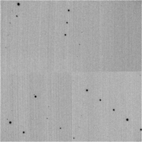
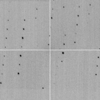
| Image | Full image | Centre region | Upper left |
| 1 |  |
 |
 |
| 31 |  |
 |
 |
Very nice spots - as expected fromm Lysozyme and a crystal that diffracts that well.
We can run autoPROC on these images with basically all defaults - only one parameter needs to be set (to tell autoPROC that the rotation axis rotates the opposite direction - this is a beamline/instrument specific setting required for autoPROC):
% process ReversePhi=yes -d 01 | tee 01.lis
This will result in a dataset
Summary data for   Project: Test Crystal: A Dataset: 0.65255

                                           Overall  InnerShell  OuterShell
---------------------------------------------------------------------------
  Low resolution limit                      28.850      28.850       1.697
  High resolution limit                      1.691       7.797       1.691

  Rmerge                                     0.019       0.028       0.036
  Ranom                                      0.017       0.042       0.040
  Rmeas (within I+/I-)                       0.024       0.059       0.057
  Rmeas (all I+ & I-)                        0.027       0.039       0.051
  Rpim  (within I+/I-)                       0.017       0.042       0.040
  Rpim  (all I+ & I-)                        0.019       0.028       0.036
  Total number of observations               20429         208         160
  Total number unique                        10397         106          84
  Mean(I)/sd(I)                               38.5        47.0        22.7
  Completeness                                95.6        95.5        73.7
  Multiplicity                                 2.0         2.0         1.9

  Anomalous completeness                      79.8        88.3        48.2
  Anomalous multiplicity                       1.0         1.0         1.0
and files:
Work in progress