Content:
Triggered by this article (Stoermer & Kenny) (thanks to J. Cole from CCDC for pointing us to this).
What do we see in the results from automatic re-refinement of those structures? The pictures were all done with Coot, 2mFo-DFc (electron density) maps at 1.0 rms and mFo-DFc (difference density) maps at 3.5 rms, respectively. Just run
  coot --pdb 6LU7_aB_refine.01_03_refine.pdb --auto 6LU7_aB_refine.01_03_refine.mtz
  coot --pdb 6Y2F_aB_refine.01_03_refine.pdb --auto 6Y2F_aB_refine.01_03_refine.mtz
after downloading the BUSTER results from here and here.
6LU7
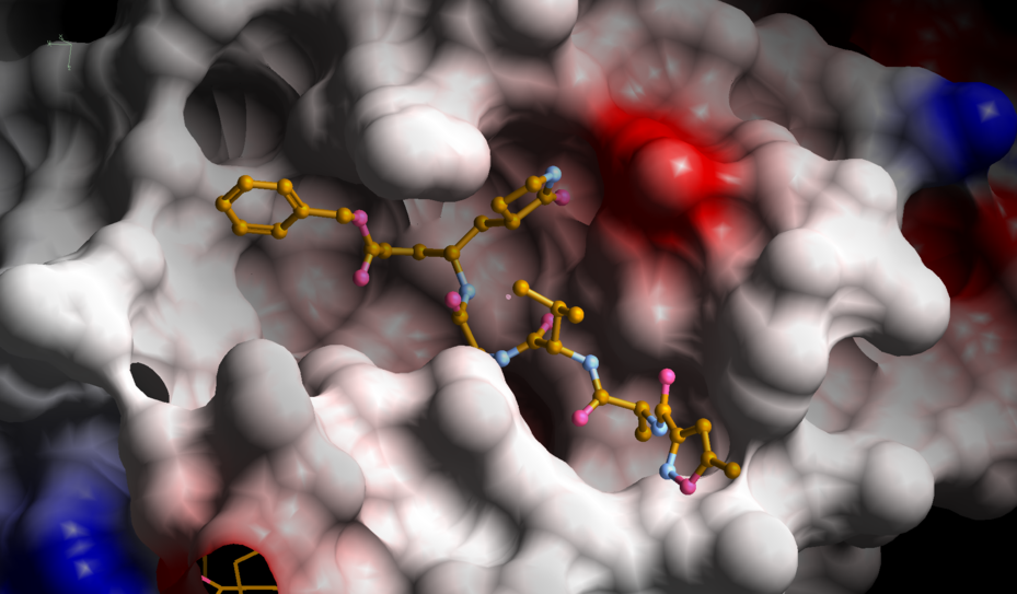 |
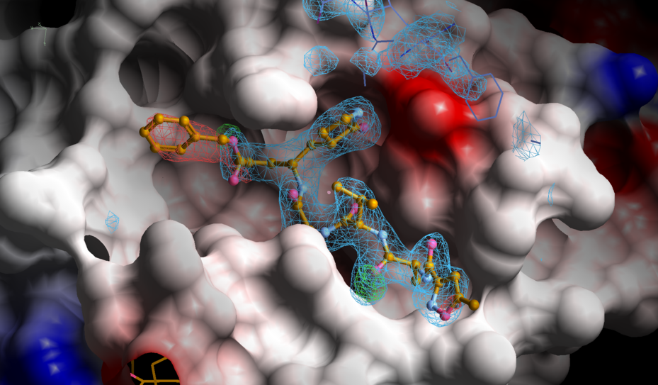 |
The difference density suggests that the phenyl group to the left is not really present in the crystal - maybe cleaved during soaking/binding or due to radiation damage? The latter is visible in negative difference density at some carboxylate groups:
 |
 |
We can try and model the probably cleaved 010 part in two ways:
| refining occupancy in BUSTER (to 0.23): |
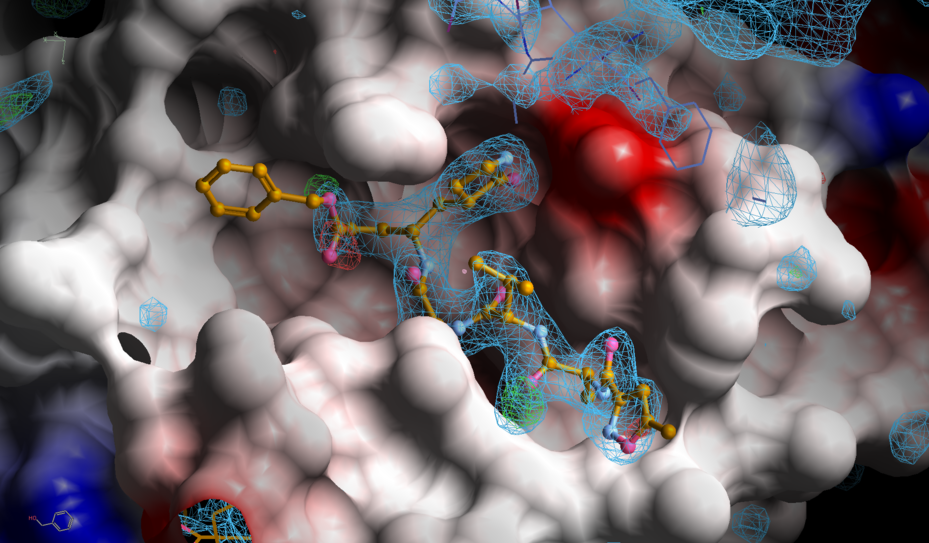 |
and
| removing 010 completely: |
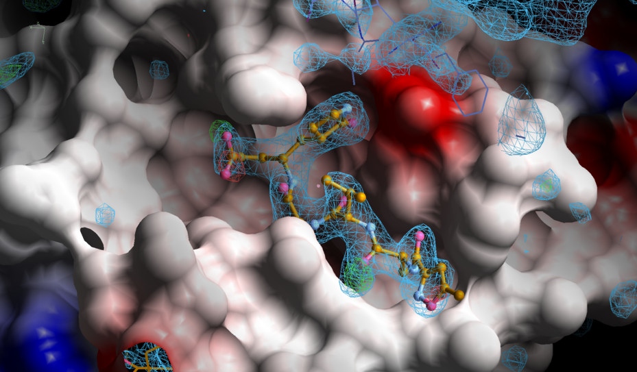 |
One has to remember that a very low occupancy can basically just represent the bulk solvent model. Since there doesn't seem to be any indication of this group being present in the difference density of the second map (where we left it completely out of the model for refinement), this does very much look like the result of cleavage.
It would be interesting to compute radiation-damage detection maps F(early)–F(late) as prepared by autoPROC analysis and computed by BUSTER. For this, the raw image data (hopefully wiht high enough multiplicity) would have to be available and (re-)processed with e.g. autoPROC. These maps could show if that part of the compound was present at the beginning of data-collection (and cleaved due to radiation) or it never made it into the crystal during soaking or co-crystallisation - an obviously very important distinction when it comes to interpretation of the structure!
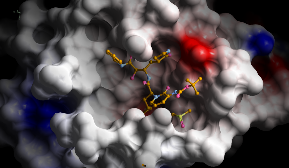
6Y2F
 |
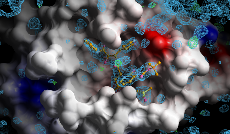 |
Nice density for the whole compound - and also several surrounding water molecules. Some indications of radiation damage in other parts of the structure:
 |